When a plaintiff accuses another party of harming them and causing them grievous injury, the burden of proof lies on that plaintiff. However, the extent of some physical injuries can’t be determined just by conducting a visual examination. In fact, some injuries affect internal organs and don’t show external visible signs at all, and the only way to see the harm suffered by the plaintiff is to take and examine medical images.
Medical images refer to pictures that visually represent the body’s interior. By using weak radiation such as X-ray or magnetic fields, internal structures can be revealed without having to cut the body open. Healthcare professionals use medical images for the following purposes:
- To collate a database of normal human anatomy that can be used to identify occurrences of abnormality
- To illustrate tissue and organ function
- To make clinical analyses, diagnose medical conditions, and treat diseases
Seeing internal injuries and ailments with our own eyes is vastly more impactful than reading about them in medical reports. To illustrate, let us say that in explaining what ails a plaintiff, a health expert uses medical jargon like “lung carcinoma due to asbestos inhalation” in their written findings. Those who are unfamiliar with the term “carcinoma” will find it difficult to understand the plaintiff’s situation, much less be able to sympathize with the injured party. Having a picture illustrate what the term actually means makes jurors and judges see what the plaintiff is going through.
Common types of medical images
Advancements in medical technology have produced numerous ways for visually representing the contents and internal processes of the human body. The most common medical images requested for court proceedings include:
X-ray images
By letting X-rays pass through the body and onto a film, bones and other internal structures — plus any foreign objects left behind during surgery — can be seen. X-ray images readily reveal bone fractures and abnormalities in the lungs, while using contrasts can provide more detail on soft tissue organs like the uterus.
CT scans
Computed tomography (CT) scanning is a more sophisticated form of X-ray imaging. Instead of being static, the X-ray transmitter and detector of a CT scanner go around the patient’s body to capture multiple images. These pictures are used to form two-dimensional slices and three-dimensional views of internal organs and structures.
CT scans have higher resolution than regular X-rays, which means that these can reveal bone damage and abnormalities in greater detail. These scans can also capture things X-rays would miss, such as tumors, internal organ injuries, internal bleeding, and brain swelling.
Related article: How law firms can save time and money by availing radiology imaging retrieval services
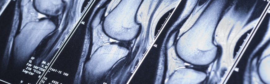
MRI images
Magnetic resonance imaging (MRI) subjects the nuclei of hydrogen molecules in the human body in a magnetic field. The molecules react by producing signals that, when detected, can form images. MRIs are commonly used to create images of the brain and spine, the cardiovascular system, and cancerous tumors.
PET images
Positron emission tomography (PET) involves introducing a radionuclide tracer in the body and detecting the gamma rays it emits. PET is often used to discover brain trauma that couldn’t be seen with CT scans and MRIs, and determine the extent of the spread of cancer in the body.
SPECT scans
Single photon emission computed tomography (SPECT) scans are similar to PET scans, except that the former can provide true 3D information. This information is often presented in cross-sectional slices that can vividly reveal damaged parts of organs, such as the brain and heart, and can even indicate injuries stemming from undiagnosed and untreated cardiac and cerebral conditions.
Obtaining and utilizing medical images in the past
It was once common practice for attorneys to mail or fax requests for radiology images. They had to wait weeks for CDs to arrive or pay exorbitant fees for overnight shipping — and pray that the CDs weren’t damaged or lost during transit. To this day, some hospitals can’t provide images any other way.
Hospitals also provide viewing software so that the image files contained in the CDs can be displayed on screens and printed as exhibits. These programs may vary from hospital to hospital and are subject to updates, which means that law firms have to manage their software to utilize medical images properly.
Opposing counsel must also be supplied with medical images, which adds a wrinkle to an already complicated process.
To persuasively present the information contained in medical images, lawyers must understand the methods used to obtain the images, as well as what the pictures actually depict. To do so, they must consult with the plaintiff’s physician or the firm’s retained medical expert. Often, they’ll have to call these medical practitioners to the stand and have them explain the images in court.
Obtaining and utilizing medical images today
Thanks to medical record and imaging retrieval services like [company_short], law firms can conveniently, inexpensively, and securely obtain medical images from healthcare providers. And if you still need to store the image files in physical media, you can use the service to save the images in CDs, too. To learn more about how we can make medical image retrieval easier for your firm, contact us today.